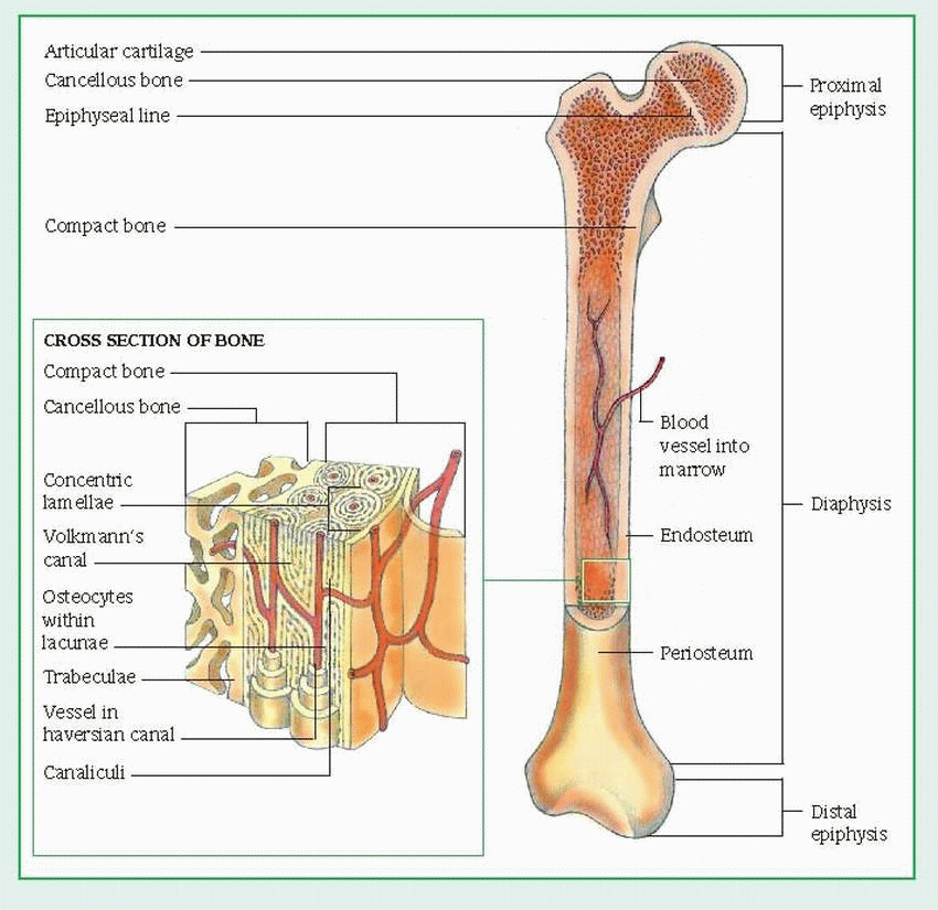
Diagram Cross Section Of A Bone / Bone Structure | Anatomy and Physiology I / Diagram with articular cartilage, marrow, spongy bone, medullary cavity, endosteum, diaphysis, and periosteum.
byAdmin-
0
Diagram Cross Section Of A Bone / Bone Structure | Anatomy and Physiology I / Diagram with articular cartilage, marrow, spongy bone, medullary cavity, endosteum, diaphysis, and periosteum.. Diagram of a cross section of the coiled cochlea. So what im going to do is jump right to the. For clarity, the cancellous bone of the metaphysis is not shown. A cross section of a human long bone. They build the entire picture, improve your understanding, consolidate the information and facilitate recall.
Patient's and clinic's names removed real brain mri slide of a young woman. Function of bone bone is a living, metabolically active and highly organized tissue consisting of a. In the last decade, considerable technological improvements have been made to repair damaged bones and tissue, such as bone cross sections with implants for microscopic examinations. (b) in this micrograph of the osteon, you can clearly see the concentric lamellae and central canals. Cross section diagrams a cross section diagram is if you would take a knife and cut through one side of a diagram to see the inside and outside in one picture.
Musculoskeletal Disorders | Basicmedical Key from basicmedicalkey.com Compact bone is the outer layer and the spongy bone forms the inner layer. Cross section of a bone diagram : In a cross section of a bone we can see two types of bone tissue: The cross section of a solid is a plane section resulting from a cut (real or imaginary) perpendicular to the length (or breadth of height) of the solid. As shown in figure 2. These bone cells (described later) cause the bone to grow, repair, and remodel throughout life. Bone is found in the shafts of long bone and consists of various cylindrical units named as haversian system 47. (micrograph provided by the regents of university of michigan.
The centroidal locations of common cross sections are well documented, so it is typically not necessary to calculate the location with the equations above.
Volume of a solid figure with uniform cross section. Explaned distal and proximal epiphysis. These bone cells (described later) cause the bone to grow, repair, and remodel throughout life. Compact bone is the outer layer and the spongy bone forms the inner layer. Patient's and clinic's names removed real brain mri slide of a young woman. For clarity, the cancellous bone of the metaphysis is not shown. Diagram of a cross section of the coiled cochlea. In the last decade, considerable technological improvements have been made to repair damaged bones and tissue, such as bone cross sections with implants for microscopic examinations. A cross section of a human long bone. Cross section diagrams a cross section diagram is if you would take a knife and cut through one side of a diagram to see the inside and outside in one picture. Spongy bone and compact bone. For example, to read this diagram literally, since the cartilage can be seen inside the. The surface features of bones vary considerably, depending on the function and location in the body.
Volume of a solid figure with uniform cross section. The surface features of bones vary considerably, depending on the function and location in the body. As shown in figure 2. Ear external and internal anatomy cross section unlabeled stock illustration 9895a hr fotosearch / wh. The centroidal locations of common cross sections are well documented, so it is typically not necessary to calculate the location with the equations above.
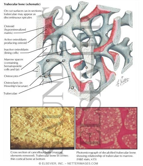
Structure of Trabecular Bone from www.netterimages.com So what im going to do is jump right to the. (b) in this micrograph of the osteon, you can clearly see the concentric lamellae and central canals. As the names suggest compact bone looks compact and the spongy bone looks like skull bone is a flat bone. Vector illustration scheme of bone cross section. Metaphseal region on the left, diaphyseal region on the right. Hope you enjoy and please. Diagram with articular cartilage, marrow, spongy bone, medullary cavity, endosteum, diaphysis, and. Ga voor hoogwaardige illustratieve kunst met een hoge resolutie naar getty images.
Cross section of a human bone.
Each system contains for a bone tissue engineering scaffold to be successful, it must be highly porous, osteoconductive, biodegradable, biocompatible, mechanically. There are trabeculae in spongy bone which gives its sponge like appearance. Cross section diagrams a cross section diagram is if you would take a knife and cut through one side of a diagram to see the inside and outside in one picture. From wikimedia commons, the free media repository. They build the entire picture, improve your understanding, consolidate the information and facilitate recall. Cochlea diagram cross section as the travellers or messenger terminals are normally interconnected, the prevalent terminal is the only a single left. Jump to navigation jump to search. For example, to read this diagram literally, since the cartilage can be seen inside the. Whereas a long bone has only one layer of compact bone (see fig 1). Diagram with articular cartilage, marrow, spongy bone, medullary cavity, endosteum, diaphysis, and periosteum. As shown in figure 2. Diagram with articular cartilage, marrow, spongy bone, medullary cavity, endosteum, diaphysis, and periosteum. Explaned distal and proximal epiphysis.
In a cross section of a bone we can see two types of bone tissue: Human respiratory system anatomical line style artistic vector illustration, medical education cross section diagram. Hope you enjoy and please. Diagram with articular cartilage, marrow, spongy bone, medullary cavity, endosteum, diaphysis, and periosteum. can be used for personal and commercial purposes. Cross section of a bone diagram :
Bone and Cartilage at University of South Florida College ... from classconnection.s3.amazonaws.com In a cross section of a bone we can see two types of bone tissue: They build the entire picture, improve your understanding, consolidate the information and facilitate recall. Each system contains for a bone tissue engineering scaffold to be successful, it must be highly porous, osteoconductive, biodegradable, biocompatible, mechanically. Volume of a solid figure with uniform cross section. Diagram of blood and nerve supply to bone. As shown in figure 2. Ear external and internal anatomy cross section unlabeled stock illustration 9895a hr fotosearch / wh. Patient's and clinic's names removed real brain mri slide of a young woman.
Patient's and clinic's names removed real brain mri slide of a young woman.
We can see there are two layers of compact bone here. Explaned distal and proximal epiphysis. As the names suggest compact bone looks compact and the spongy bone looks like skull bone is a flat bone. Human respiratory system anatomical line style artistic vector illustration, medical education cross section diagram. These bone cells (described later) cause the bone to grow, repair, and remodel throughout life. (micrograph provided by the regents of university of michigan. Diagram with articular cartilage, marrow, spongy bone, medullary cavity, endosteum, diaphysis, and periosteum. The centroidal locations of common cross sections are well documented, so it is typically not necessary to calculate the location with the equations above. For example, to read this diagram literally, since the cartilage can be seen inside the. A cross section of a human long bone. Cross section of a human bone. In a cross section of a bone we can see two types of bone tissue: Vector illustration scheme of bone cross section.
For example, to read this diagram literally, since the cartilage can be seen inside the cutaway section of bone, it incorrectly indicates that the cartilage in fact goes through the bone structure, rather than just being found around the bone end cross section of a bone. The centroidal distance , c , is the distance from the centroid of a cross section to the extreme fiber.